Novel laser-fabricated plasmonic chip combines 185 nm Raman resolution and deep learning to identify and sort cancer cells with 96.2% accuracy
CHENGDU, SICHUAN, CHINA, September 9, 2026 /EINPresswire.com/ — Accurate and efficient isolation and recovery of interest cells allow detailed analyses of cellular function, growth and culture, and gene-expression profiling of cells within heterogeneous populations. Researchers have developed a novel microfluidic surface-enhanced Raman scattering (SERS) chip capable of performing deep-learning-assisted, label-free single-cell identification and sorting. The results could be used to guide future research on the design and development of non-invasive, efficient cell-sorting techniques with potential applications in clinical diagnostics and precision medicine.
Cell sorting is a basic yet vital procedure in precision biomedicine. Cell sorting separates mixed cells into pure subgroups based on their unique features. Biological tissues and blood contain diverse cell types, and mixed samples blur true cell signals in gene or protein tests. By sorting, researchers can get uniform cells to study cell functions, stem cell development, and disease mechanisms clearly. It also supports drug screening and cell product quality control. Without these techniques, accurate single-cell analysis and modern cell-based treatments would be impossible. The analysis of rare cells, such as cancer stem cells and circulating tumor cells, to assess their functional responses is rapidly gaining prominence given its relevance for its potential to address issues in the development of new drugs and clinical diagnostics.
Traditional flow-cytometry-based sorting was first described in the 1960s. Now, the most widely used methods are fluorescence-activated cell sorting and magnetic-activated cell sorting. They pass single cells through laser detection; charges target cell droplets and deflect them via electric fields for collection. Recently, to avoid labeling and invasive manipulation, researchers have developed several types of label-free cell sorters, including image-activated cell sorting (IACS), acoustics-activated cell sorting (AACS), and Raman-activated cell sorting. These systems utilize intrinsic cellular properties, such as geometric morphology, acoustic characteristics, and molecular composition measured by Raman spectra to achieve identification and sorting, respectively. However, these techniques exhibit limited sorting accuracy in complex cell populations.
To improve the identification accuracy, deep-learning approaches based on neural networks (NNs) have recently been developed for cell sorting. However, NN algorithms are computationally complex and often unsuitable for use by untrained users and the primary barriers to label-free cell sorting are low sensitivity and limited spatial resolution, because these parameters are directly related to the accuracy of the entire identification-to-isolation workflow.
Surface-enhanced Raman scattering (SERS), which provides an effective and non-invasive approach for identifying single-molecule fingerprints in biosensing, offers ultra-sensitive capabilities for trace detection and analysis. Thanks to the high accuracy of Raman signals and short acquisition time, SERS can facilitate cell sorting because Raman analysis requires less time compared with IACS and AACS. In addition, the Raman mapping provides spatially resolved Raman signals, thereby reducing the total processing time. In recent years, deep-learning-based SERS techniques have been developed for cell identification. Previous studies used Au nanoparticles as SERS substrates to collect cancer-cell signals and applied the resulting data to NN-based deep-learning models for early cancer diagnosis. However, appropriate selection of Raman tags or biomarkers is critical for accurate SERS analysis, even when using deep-learning models, and it is important to consider that deep learning has limited utility as a universal technology for cell identification.
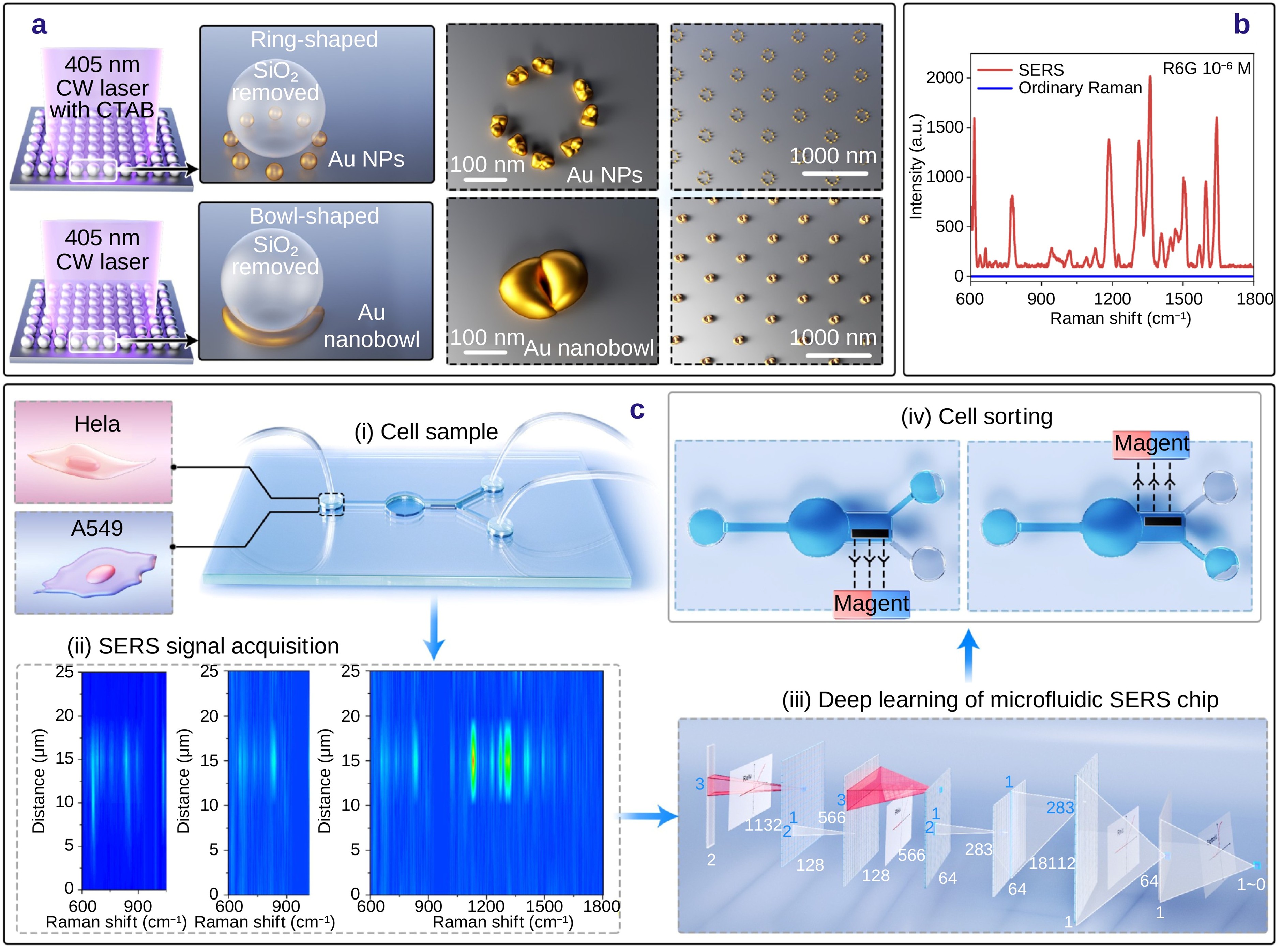
Recently, the research group of Professor Koji Sugioka and Dr. Shi Bai from Advanced Laser Processing Research Team, RIKEN, proposed a new approach to fabricate a SERS substrate with uniformly distributed hotspots by laser near-field reduction of Au ions. Their work is made available online on July 29, 2026, in the Early View section of Opto-Electronic Advances.
“This technique allows the creation of plasmonic ring-shaped nanostructure arrays by introducing cetyltrimethylammonium bromide into the precursor solution,” explained Prof. Sugioka. The Au nanoparticle has a size of ~28 nm. Meanwhile, the number of Au nanoparticles and their gaps in a ring structure are related to the reduction time, which is critical for robust SERS performances.
Raman mapping indicates that the ring-shaped nanostructure arrays show ultrahigh spatially uniform Raman enhancement with a relative standard deviation of 2%. The high spatial resolution (~185 nm) of SERS analysis was also experimentally confirmed by Raman mapping. Significantly, it was found that the spatial resolution is essential for deep-learning label-free cell identification. The results showed owing to the high spatial resolution of ring-shaped nanostructure array, the accuracy of cell identification increased from 75% to 96.2%, illustrating that spatial resolution was a key factor and should be considered in future related studies.
As a proof of concept, the researchers fabricated a cell sorting device by femtosecond laser processing. The cancer or non-cancer cells were successfully sorted in a “Y”-shaped microchannel. “The preliminary results indicate that the developed sorter achieves precise cell sorting and separation without the need for biomarker or Raman-tag labeling,” highlighted Prof. Sugioka.
Reference
Title of original paper: Deep-learning-assisted label-free single-cell identification and sorting using femtosecond laser fabricated microfluidic surface-enhanced Raman scattering (SERS) chips with plasmonic ring-shaped nanostructure arrays
Journal: Opto-Electronic Advances
DOI: https://doi.org/10.29026/oea.2026.260071
About Opto-Electronic Advances
Launched in March 2018, Opto-Electronic Advances (OEA) is an open-access, peer-reviewed SCI journal. Indexed in major databases including SCI, EI, and Scopus, it is guided by an international editorial board spanning 17 countries. OEA provides a fast-track platform for high-impact empirical and theoretical research across optics, photonics, and optoelectronics. Key focus areas include light sources, sensors, nanophotonics, plasmonics, biophotonics, optical imaging, intelligent optics, and optical communications, catering to researchers, academicians, and industry professionals seeking cutting-edge developments in light-driven technologies.
Website: https://www.oejournal.org/oea/en/
About Professor Koji Sugioka from the RIKEN Center for Advanced Photonics, Japan
Prof. Dr. Koji Sugioka received his B. S., Ms. Eng., and Dr. Eng. Degrees in electronics from Waseda University (Japan) in 1984, 1986, 1993, respectively. He Joined RIKEN in 1986 and is currently a Team Director of Advanced Laser Processing Research Team at RIKEN Center for Advanced Photonics. He was awarded the degree of Doctor Honoris Causa from University of Szeged, Hungary in 2018. He is currently a board of trustee member of the Laser Institute of America (LIA) and a member of board of directors of the Intl. Academy of Photonics and Laser Engineering (IAPLE), the Japanese Laser Processing Society, and the Japan Society of Laser Technology. He is a Fellow of SPIE, Optica, LIA, IAPLE, and JSAP. He is also a former editor-in-chief of Journal of the Laser Micro/Nanoengineering (JLMN) and currently an editor of Nanomaterials, International Journal of Extreme Manufacturing (IJEM), and Opto-Electronic Advances (OEA).
About Dr. Shi Bai from the RIKEN Center for Advanced Photonics, Japan
Dr. Shi Bai received his Ph.D. in optical engineering from Beijing University of Technology in 2018 and attended Prof. Sugioka’s team in 2019 as a special postdoctoral researcher, visiting researcher in 2022, and research scientist in 2024. Now, he is working at Shanghai Institute of Optics and Fine Mechanics, CAS, as a researcher. His research activities are focused on the ultrafast laser nanofabrication and ultra-sensitive SERS microfluidic chips and the applications in environments and biomedicine.
Funding information
Anming Hu acknowledges support from a research grant funded by the Institute of Security & Sustainability at the University of Tennessee Knoxville, the USA. This study was funded by the National Natural Science Foundation of China (Grant No. 52375422) and Science Research Project of Hebei Education Department, China (Grant No. BJK2024021).
Siyi Ma
Editorial Office of Opto-Electronic Journals Group, address
email us here
Legal Disclaimer:
EIN Presswire provides this news content “as is” without warranty of any kind. We do not accept any responsibility or liability
for the accuracy, content, images, videos, licenses, completeness, legality, or reliability of the information contained in this
article. If you have any complaints or copyright issues related to this article, kindly contact the author above.

Media gallery